Instant Download
Your unique download link appears right after you complete your purchase, and is also sent instantly to the email address you provided.
Couldn't load pickup availability

Your unique download link appears right after you complete your purchase, and is also sent instantly to the email address you provided.
Yes
Diagnostic-Imaging
pediatric cardiologists, sonographers, and obstetricians
Mixed (Video + PDF)
Mixed (Video + PDF)
Other
1. Overview
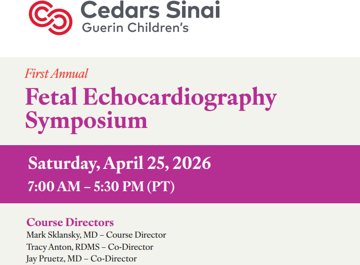
The Cedars Sinai 1st Annual Cedars-Sinai Fetal Echocardiography Symposium provides a comprehensive update on the latest clinical guidelines and practices in prenatal cardiac imaging. The program focuses on advanced diagnostic techniques, the early detection of complex congenital heart defects, and multidisciplinary strategies to optimize perinatal outcomes and maternal counseling.
Our entirely refreshed 2026 fetal echocardiography symposium remains true to the mission of providing a single day full of clinical tips and pearls for everyone involved with fetal cardiac imaging in low-risk or high-risk pregnancies. This year’s course, revamped with an expanded faculty of expert clinicians and teachers, includes didactic lectures by world-class sonographers, maternal fetal medicine and pediatric cardiology faculty, two round-table multidisciplinary discussions, impromptu testimonials from young patients with major forms of congenital heart disease, question and answer sessions with participation from the live and live-streamed audiences, and a hands-on workshop for participants to scan actual patients towards the end of the afternoon.
2. Learning Objectives
Discuss current guidelines for fetal cardiac screening and fetal echocardiography
Improve the basic approach to screening low risk pregnancies for fetal heart disease.
Identify new ways to optimize image quality and angle of acquisition.
Understand the optimal use of color flow Doppler in the evaluation of the fetal heart.
Learn to acquire high quality images clips of the four chamber view, outflow tracts, and three vessel views.
Evaluate and diagnose major forms of congenital heart disease, fetal cardiac tumors, and ventricular dysfunction/cardiomyopathy
Learn to collaborate across interdisciplinary teams to develop coordinated prenatal to postnatal care plans that optimize outcomes for infants with congenital heart disease.
3. Target Audience
Best for pediatric cardiologists, sonographers, and obstetricians who want advanced updates on fetal echocardiography and congenital heart disease.
4. Topics
2026 Guidelines for Fetal Cardiac Imaging- Screening vs Fetal Echocardiography
Fetal Cardiomyopathy and Cardiac Tumors- Importance and Challenge of Early Detection
Fetal Detection-Evaluation of Tricuspid Valve Disease- Lessons Learned
Impact of Delayed Detection of Congenital Heart Disease- The Adult Congenital Cardiologist’s Perspective
Introduction and Welcome- Dr. Sklansky
Making the Diagnosis of TGA-TOF-DORV- Keys to Prenatal Detection-Evaluation
Pioneer Lecture- The Future of Fetal Cardiac Imaging
Right Ventricular Outflow Tract Obstruction (PS and PA-IVS)- Prenatal Detection-Evaluation
Round Table Discussion- How to Train Your Office for Fetal Cardiac Imaging
Secrets to Optimizing 2D Image Quality and Angle of Acquisition
Spectral and Color Doppler in the Fetal Cardiac Evaluation- Clinical Pearls
TAPVR and Coarctation of the Aorta- Challenge and Secrets of Prenatal Detection
The Three-Vessel View- From Basic to Advanced Evaluation
Why It Matters- Prenatal Consultation with a Congenital Heart Surgeon